Table of Contents
Histology studies the composition and structure of plant and animal tissues. The usage of terms histology and microscopic anatomy are sometimes colloquial. Histology’s goal is to determine the organization of tissues. Let’s learn more about histology and its uses.
History of histology
What is histology? How did it evolve? The microscope and histology have been around for centuries. Robert Hooke published ‘ Micrographia’ in 1665, which was one of the first significant milestones in this field.
It is the first published instance of the term ‘cell.’ Hooke compared the walled plant cells he was studying to a monk’s quarters.
What is histology?

The scientific study of the microscopic structure (microanatomy) of cells and tissues is known as histology. The term ‘histology’ derives from the Greek words ‘histos’ (tissue or columns) and ‘logia’ (study). The term ‘histology’ first appeared in a book written in 1819 by German anatomist and physiologist Karl Meyer. You can trace back its origin to 17th-century microscopic studies of biological structures performed by Italian physician Marcello Malpighi.
How does histology work?
Histology courses focus on preparing it’s slides, which requires prior knowledge of anatomy and physiology. One typically teaches light and electron microscopy techniques separately.
The five steps to preparing slides are as follows
- Fixing
- Processing
- Embedding
- Sectioning
- Staining
You must first fix cells and tissues to prevent decay or degradation. Processing is required to avoid excessive tissue alteration. Cutting small samples into thin sections is suitable for microscopy by embedding them within a supporting material. Microtomes and ultramicrotomes are special blades used for sectioning. Cells are stained and mounted on microscope slides.
The most common stain is a mixture of hematoxylin and eosin (H&E stain). Cellular nuclei are stained blue with hematoxylin, while the cytoplasm is stained pink with eosin. Images of H&E slides are typically pink and blue. Toluidine blue stains the nucleus and cytoplasm blue, but mast cells purple. Wright’s stain turns red blood cells blue/purple while changing the color of white blood cells and platelets.
Types of tissues
Plant tissue and animal tissue are the two broad categories of tissues. To avoid misunderstanding, ‘plant anatomy’ is the common name for plant histology. The following are the main types of plant tissues-
- Vascular tissue
- Dermal tissue
- Meristematic tissue
- Ground tissue
One can classify all tissue in humans and other animals into one of four groups
- Nervous tissue- Cells (neurons and glial cells) and extracellular matrix make up nervous tissue. The ECM of nervous tissue is composed primarily of ground substance, with few to no protein fibers.
- Muscle tissue- Muscle tissue continues to synthesize and contract. Their classification includes either skeletal, cardiac, or smooth. These are either voluntary (skeletal) or involuntary based on their functional properties (cardiac and smooth muscle).
- Epithelial tissue- Epithelial tissue can cover external surfaces (such as skin), line the inside of hollow organs (such as the intestines), and form glands. It comprises tightly packed epithelial cells with minimal extracellular matrix (ECM). The cell arrangement is on top of the basement membrane, which contains dense irregular connective tissue (BM).
- Connective tissue- Connective tissue connects, separates, and upholds the body’s organs. It comprises a few cells and a lot of extracellular matrix. The ECM contains different protein fibers (collagen, reticular, elastic) embedded in ground substance.
Subcategories of these main types include epithelium, mesothelium, mesenchyme, germ cells, and stem cells. You can also use histology to study microorganisms, fungi, and algae structures.
Career in histology
The following are some of the careers
Histologist

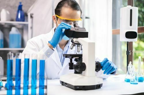
A histologist is someone who prepares tissues for sectioning, cuts them, stains them, and images them. Histologists work in labs and have highly refined skills, such as determining the best way to cut a sample, staining sections to reveal essential structures, and imaging slides using microscopy. Biomedical scientists, medical technicians, histology technicians (HT), and histology technologists work in a this lab (HTL).
Pathologist
Pathologists examine the slides and images created by histologists. Pathologists specialize in identifying abnormal cells and tissues. Many conditions and diseases, including cancer and parasitic infection, can be identified by a pathologist, allowing other doctors, veterinarians, and botanists to devise treatment plans or determine whether an abnormality caused death.
Histopathologists
Histopathologists are experts in the study of diseased tissue. A medical degree or doctorate is typically necessary for a career in histopathology. Many scientists in this field hold dual degrees.
Use of histology
Histology is essential in science education, applied science, and medicine.
Biologists and medical students
It is essential to teach histology to biologists, medical students, and veterinary students because it helps them understand and recognize different types of tissues. Histology, on the other hand, bridges the gap between anatomy and physiology by demonstrating what happens to tissues at the cellular level.
Archaeologist and Paleontologist
Histology is a technique used by archaeologists to study biological material recovered from archaeological sites. The most likely sources of information are bones and teeth. Paleontologists can salvage valuable material from organisms preserved in amber or frozen in permafrost.
Diagonizes
One of the purposes of histology is to diagnose human, animal, and plant diseases and to analyze treatment effects.
Autopsies
Autopsies and forensic investigations use histology to help explain anomalous deaths. In some cases, microscopic tissue analysis can reveal the cause of death. In some instances, microanatomy may disclose information about the environment after death.
Key takeaways
- Histology courses emphasize the preparation of its slides, which necessitates prior knowledge of anatomy and physiology.
- Plant histology is commonly known as plant anatomy to avoid misunderstanding.
- Histology has various career opportunities, such as histologist, pathologist, and histopathologist.
Did you find this blog informative? If so, please share your thoughts in the comments section below. Click here to contact us for more information on histology. We would be happy to assist you with your queries.
Liked this blog? Read next: Smallest continent on the planet- Australia
FAQs
Q1. What are the three main types of connective tissue?
Answer- Loose connective tissue, dense connective tissue, and specialized connective tissue are the three main types of connective tissue.
Q2. Who is the father of histology?
Answer- Marie François Xavier Bichat was a French anatomist and pathologist known as the ‘Father of Modern Histology.’
Q3. What are the types of nervous tissue?
Answer- Our nervous tissue comprises three major parts- nerves, the spinal cord, and the brain.


